 Health care has evolved gradually and consistently with the emergence of new technologies, especially those designed to improve the quality of patient care. New technologies long have aided clinicians in diagnosing and treating patients, particularly those with complex medical issues. This level of complexity is no more evident than in a health care facility's imaging department.
Within the past 20 years, combining imaging technologies has become popular. This practice, in fact, has become common in the world of nuclear medicine, where both positron emission tomography (PET) and single-photon emission computed tomography (SPECT) have been combined with computed tomography (CT) to create PET/CT and SPECT/CT, respectively.
Until recently, most clinicians have been leaning on PET/CT and SPECT/CT for the majority of their work in oncology. But that's about to change.
What is PET/MR?
PET/MR combines PET, which uses a tracer to highlight tumors, and magnetic resonance (MR) imaging, which uses a strong magnetic field to produce images of soft-tissue filled areas. The advantage of combining these two technologies into one system is that oncologists now are able to get the best available image of a tumor located in a soft tissue area.
This hybrid system can provide clinicians with new insights into the worlds of neuroscience and neurological disorders. It also will allow oncologists the option to scan a patient's whole body, which is especially important with increasing instances of cardiovascular disease and cancer. Imagine a system that can scan a patient's entire body with the sensitivity to detect any brain, bone or liver tumors all in one scan.
Another advantage of PET/MR is the ability to utilize other imaging techniques to complement the main technology. For example, by using spectroscopy with a PET/MR scan, clinicians can investigate the properties of specific organs to determine if there are any abnormalities. Functional MRI (fMRI) also can be used along with PET/MR to assess brain function by detecting a contrast dependent on the blood-oxygenation level. Combining PET with cardiac MRI and whole-body MR angiography also enables physicians to detect and differentiate between vulnerable plaques.
Along with these advantages, PET/MR also eliminates the existence of ionizing radiation incurred by the patient when compared with PET/CT. The amount of radiation exposure through PET/CT can be significant, especially if repeated whole-body examinations are required for monitoring purposes. By using PET/MR instead of PET/CT, radiation is reduced to the minimal amount emitted by the radiopharmaceutical required for the PET portion of the scan.
Present and future
The first PET/MR prototype was assembled at the University of California–Davis in 1997. Since then, the major imaging vendors have been working to perfect the PET/MR technology to serve the needs of health care.
As of 2011, the first two PET/MR models were approved for sale in the United States by the Food and Drug Administration. One additional model since has been approved.
In general, there are two main configurations in PET/MR.
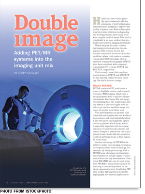
• The simplest configuration is a tandem one, where PET and MR images are taken sequentially in two separate scanners. This can be achieved either by having both systems in the same room or in separate rooms. The former rotates the patient table between imaging systems, while the latter moves the same patient transfer tabletop between both rooms and docks it on both system gantries. In addition to the simplicity of image registration, the advantages of a tandem configuration are that it's less expensive than a concurrent system and it minimizes claustrophobia because of the large space between scanners.
• The more complex method, known as concurrent PET/MR imaging, combines both PET and MR in a single platform. The main advantage is a better overall image, due to the simultaneous generation of images. When both PET and MR images are taken concurrently, there is no potential for an organ to move between images and disrupt the quality of the image. Another advantage of capturing both images simultaneously is that highly quantitative measurements can occur, the most important being PET/fMRI. However, these systems typically are more expensive than tandem systems.
The future of PET/MR systems will depend on how user-friendly the systems are and how reproducible the multimodality images become. There was a quantum leap in technology development with the emergence of the concurrent PET/MR system. This addressed the major shortcomings with the tandem systems, including issues with patient movement and the need for reimaging of patients. For these reasons, it is likely that the concurrent PET/MR system will become the more popular PET/MR technology for facilities.
Effects on facilities
So, what is holding health care facilities back from jumping at the chance to purchase a PET/MR system? With all the advantages over PET/CT, including safety and better image detail, why are administrators being so cautious?
Purchasing. For starters, a PET/MR system costs approximately $7 million, according to Plymouth Meeting, Pa.-based ECRI Institute's PricePaid database. This includes the PET and MR software, the necessary coils, the majority of the workstation components, installation and training costs, and universal power supply components. While the price may not seem too bad for a new and emerging technology, it doesn't include construction or renovation costs, which will be necessary before installation can take place.
Construction. Not many facilities have open space large enough to house this new system without renovations. Moreover, existing PET and MR spaces are not equipped to handle the necessary requirements for the other technology. For example, an existing PET room will not have the structural support in the floor to support the MR magnet without reinforcement, while an existing MR space will not have the additional support areas necessary for PET studies.
A larger room is necessary because the typical PET or MR room size will not suffice for either a tandem or concurrent system. The standard room size required for a tandem configuration PET/MR system is 60 square meters, whereas a concurrent PET/MR system requires a minimum of 33 square meters of space. Because these systems use both PET and MR technologies, shielding for magnetic fields, radio frequency and radiation is required for the entire suite. This means a total renovation of the available space and a considerable amount of testing and environmental analysis to ensure that the diagnostic procedures being performed do not affect the rest of the hospital.
Any health care facility planning a PET/MR suite also needs to take into account the layout and flow of patients before construction begins. A single scanner will need two to three uptake rooms, where patients can wait after they are injected with the radioactive tracer.
Typically, patients wait in these rooms for approximately an hour while the tracer accumulates in any body tissue that may be cancerous. PET/MR patients also need a dedicated toilet or a "hot toilet" that is shielded and in close proximity to the uptake rooms. It is recommended that these patients not walk long distances throughout the facility because of the possibility of the radiopharmaceuticals affecting sensitive instruments.
At the end of this process, patients require a cooldown period, which means a cooldown room is also necessary.
Along with the additional patient support space requirements, a PET/MR imaging suite also must comply with the American College of Radiology's four-zone layout requirements necessary for any space with the potential for magnetic field hazards.
Essentially, the layout conceptually divides a suite into four zones. Zone 1 is considered to be any area that is freely accessible to the general public and outside of the PET/MR environment itself (e.g., dressing rooms and waiting rooms). Zone 2 is the interface between Zone 1 and the strictly controlled Zones 3 and 4. Zone 2, in this case, includes the uptake rooms, while Zone 3 encompasses any controlled access areas that have the potential to be hazardous to patients, including magnetic field hazards outside of the actual MR room. An example of a Zone 3 area would be the equipment control room looking in toward the imaging system. Finally, Zone 4 is the area in which the scanner magnet is located and needs to be clearly marked as such.
These requirements must be considered for either tandem or concurrent systems. Installation of a PET/MR system takes considerable preconstruction planning to ensure good workflow, and a well-thought-out renovation also will optimize the facility's patient flow. Whether a facility decides to retrofit an existing area or build a completely new space, the appropriate personnel should be consulted before beginning this major undertaking.
New challenges
PET/MR systems combine two technologies with a long list of requirements for facilities. Although they offer many advantages, the site-planning considerations can be daunting.
While all of the variables make the planning and installation of PET/MR a challenge, it is attainable with the right personnel involved in the decision-making process. HFM
Scott DiDonato is an associate in the applied solutions group at ECRI Institute, Plymouth Meeting, Pa. He can be reached at sdidonato@ecri.org.
| Sidebar - Safety issues when installing PET/MR |
| Any time imaging modalities are involved, patient safety is an issue. Add to this the fact that positron emission tomography/magnetic resonance (PET/MR) imaging has radiopharmaceutical and magnetic field hazards, and this technology now moves to the forefront of safety officers' minds. It is imperative that clinicians put policies in place to prevent safety problems. It is also necessary that clinicians stress the importance of safety both to the technicians and the patients. When performing a PET scan, the patient receives a radiopharmaceutical that requires an hour to be absorbed fully by the body. This requires patients to sit and wait before they are scanned. During this lengthy wait time, patients and their belongings will stay in the suite's uptake rooms. It is imperative that once this hour is complete, a patient's belongings remain in the uptake rooms until the scan is finished. If not, objects and personal belongings have the potential to become hazards to patients. It is recommended that a facility invest in ferromagnetic detectors — wall hung and handheld — and put a policy in place to screen all patients prior to scanning. Safety precautions also need to take place when dealing with additional equipment in the scan room. Nuclear medicine clinicians and technicians do not need to worry about the equipment present during PET/computed tomography scanning. However, by adding MR to PET, everything in the room must now be MR-compatible. This includes the vital signs monitor, contrast injector and anything else that may be magnetic. It is important to consult with MR safety personnel to ensure that all equipment is MR-safe and will not harm the patient. In general, PET/MR is a new technology likely to be placed in a nuclear medicine department of a hospital. It is important to review the safety procedures and have policies in place to ensure a patient's safety when being scanned by this powerful technology. |